
The Disturbing World of Primitive Brain Surgery ,Without the aid of modern anesthesia or, for that matter, modern disinfection, some Bronze Age practitioner brandished an instrument with a sharp, beveled edge and began a gruesome business.
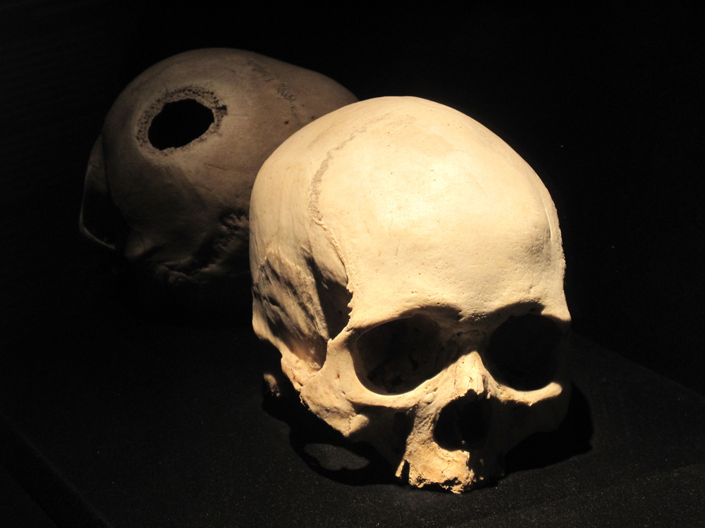
Around 1500 BC, a primitive surgeon cut a polygon from the scalp just above the left eye and peeled it off, leaving scratches in the bone below. Then began the difficult process of cutting the “living bone”, according to new archaeological surveyleaving remarkably clean furrows and gouging segments that are finally removed using “leverage”.
But don’t worry: researchers believe the inch-wide hole hasn’t stopped short of breaching the defense membrane of the dura mater which surrounds the brain.
Read more: The earliest evidence of amputation is 31,000 years old
Ancient surgery
This dramatic process, a type of trephination or craniotomy, still occurs today, albeit in a much different form – to reduce brain swelling and respond to other emergencies. In the ancient world, would-be surgeons sometimes used primitive drills or saws on humans and even on a child, with brain injuries, scurvy or intracranial infectionsometimes it kills them.
“We have evidence that trepanation has been this universal, widespread type of surgery for thousands of years,” says Rachel Kalisher in a press releasePhD candidate at Brown University and lead author of the study.
The new case stood out, in part, because of its location in the Middle East, where trepanations rarely appear in the archaeological record, and because the operation, a medical Hail Mary, lacked a clear impetus.
Procedure for the ancient elite
Researchers discovered the man’s burial site beneath a residential home in the once-prosperous city of Megiddo, in what is now modern Israel. a very battle-driven city on the important trade road Via Maris.
They found bone fragments from the procedure, suggesting that the patient or someone else placed them back into the wound to aid healing. Investigators also found food offerings and fine pottery, as well as the bones of the man’s brother, indications that the two had elite status in the nearby palace. Local residents buried the two in an earthen pit near another, larger tomb beneath the building, filled with 17 bodies adorned with gold, silver and bronze jewelry and fine bone inlay.
The brother who received the trephination died soon after, the study concluded, because the bones showed no signs of healing. Many other bones belonging to the man showed a different type of damage, telltale gouges and “penciling” that made them look like weathered wood.
What was it and what is its connection to Bronze Age brain surgery?
An ancient disease
The researchers searched for answers and suggested that perhaps the brothers had suffered lesions from a years-long infection such as osteomyelitis, syphilis, tuberculosis or leprosy. Of these possibilities, leprosy stood out because it is often shared between family members who must remain in close contact for months in order to transmit the disease. The ancient disease attacks the skin, the peripheral nervous system and even the bones themselves.
Read more: Ancient medical treatments that are still used today
Although leprosy does not directly affect the brain, it can cause neuropathic pain, a burning sensation, blindness, and the characteristic sores and swellings, including on the face. Although one of them may have triggered the operation, Kalisher is still looking for answers. With the help of the Max Planck Institute for Evolutionary Anthropology, she is now testing bone lesions for leprosy DNA.
If successful, she will identify one of the world’s earliest cases of leprosy and come closer to explaining the trepanation at Megiddo.
“You have to be in a pretty awful place to have a hole drilled into your head,” she says in a press release.
The unnamed patient may also have suffered from a number of congenital conditions that somehow precipitated the surgery, the study suggests, due to the presence of a relatively common cranial “seam” or furrow and the presence of an extra molar in his jaw. But the study could not confirm such a condition.